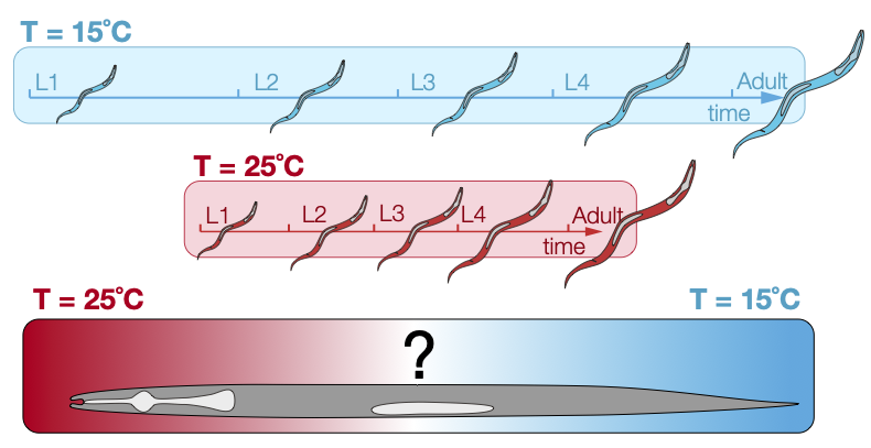
Temperature impacts all biochemical reactions inside a cell. For developing multicellular organisms, temperature fluctuations pose challenges because morphogenetic events depend on both, spatially and temporally coordinated cellular decisions. Despite this, most multicellular systems show a surprising degree of robustness with respect to temperature changes within certain temperature limits. With climate change pushing organisms more and more often towards those limits, it is important to understand how those limits are set, what molecular mechanisms coordinate overall development of an organism and whether there are ways to control these mechanisms. To address this question, our team has developed a new experimental setup, that allows us to place a small multicellular model organism (the nematode roundworm C. elegans) in a linear temperature gradient during its larval development. In our setup, the anterior part (head) of the animal can be maintained at either warmer or colder temperatures compared to the posterior part of the animal while high-resolution live-imaging is performed for several days. Surprisingly, animals develop normally in these extreme conditions, suggesting intercellular and interorgan signaling to achieve and maintain developmental coordination (Schlang et al., in preparation). Similar temperature gradient experiments have been performed with embryos of fruitflies1 [1] and frogs2[2], but gave strikingly different results, implying novel molecular mechanism underlying developmental synchrony during the post-embryonic phase of development.
The goal of this internship is to perform the first characterization of molecular players that may mediate tissue synchrony with focus on the C. elegans hypodermal stem cells. Using fluorescent reporters for developmental genes and mutants for candidate genes in the insulin and serotonin signaling pathways, the intern will investigate what may help the animal counteract the effects of this extreme environmental condition. Do to so, the student will perform spinning-disc confocal live-microscopy of animals kept in microfluidics device, an analyze various gene expression and cell division synchrony with and without the temperature gradient and how this may be changing in various genetic backgrounds. Additionally, we have recently developed technologies that allow us to visualize and analyze transcription dynamics in developing larva in real-time using the MS2-MCP-GFP tethering system3 [3]. The student will use this system to ask whether and how transcriptional kinetics of developmental genes are affected at different temperatures and how the temperature gradient condition impacts these kinetics.
- Dynamics of Drosophila embryonic patterning network perturbed in space and time using microfluidics. Nature (2005) Lucchetta et al. https://doi.org/10.1038/nature03509 ↩︎
- Desynchronizing Embryonic Cell Division Waves Reveals the Robustness of Xenopus laevis Development. Cell Reports (2017) Anderson et al.. https://doi.org/10.1016/j.celrep.2017.09.017 ↩︎
- Circadian rhythm orthologs drive pulses of heterochronic miRNA transcription in C. elegans. bioRxiv (2022), in revision; Kinney B*, Sahu S*, Stec N, Hills-Muckey K, Adams DW, Wang J, Jaremako M, Joshua-Tor L, Keil W*, Hammell CM*; https://doi.org/10.1101/2022.09.26.509508 ↩︎
Team References
- Circadian rhythm orthologs drive pulses of heterochronic miRNA transcription in C. elegans. Developmental Cell (2023), Kinney B*, Sahu S*, et al.; https://doi.org/10.1101/2022.09.26.509508
- An Epigenetic Priming Mechanism Mediated by Nutrient Sensing Regulates Transcriptional Output during C. elegans Development. Current Biology (2021) Stec N, et al.; https://doi.org/10.1016/j.cub.2020.11.060
- HLH-2/E2A Expression Links Stochastic and Deterministic Elements of a Cell Fate Decision during C. elegans Gonadogenesis. Current Biology (2019) Attner MA*, Keil W*, et al. ; https://doi.org/10.1016/j.cub.2019.07.062
- Long-Term High-Resolution Imaging of Developing C. elegans Larvae with Microfluidics. Dev Cell (2017) Keil W, et al. ; https://doi.org/10.1016/j.devcel.2016.11.022
